OCT Scanning

Optical Coherence Tomography — Woy Woy, NSW — Peninsula Eyecare Izzards Optometrists

What Is an OCT Scan?

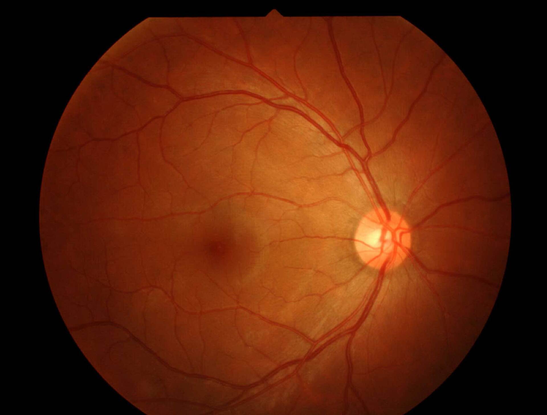
Optical coherence tomography (OCT) is a non-invasive imaging technology used to accurately identify potential eye issues by taking high-resolution cross-sectional images of the retina. OCT is similar to ultrasound testing, except that the imaging measures light rather than sound. It is a way for us to inspect the back of the eye, including the macula, optic nerve, retina, and choroid.


We can view the back of the eye during an eye examination, but sometimes we need to inspect further or capture more detail below the surface, which standard techniques don’t do. In some ways, it’s comparable to doing an "MRI of the retina." Some describe it as an optical ultrasound because it images reflections between tissues to provide doctors with cross-sectional images.


Optical coherence tomography works by using interferometry, enabling eye care professionals to image tissue with light instead of X-rays, sound or radio frequency. It works by shining a beam of light into the eye. Different tissues reflect part of this light, and images are built based on this internal reflection differential.


An OCT images to approximately 2-3 mm below the surface of the tissue. Images are obtained clearly through a transparent window, such as the cornea. The light that emits into the eye is safe. The output is near-infrared light so that no eye damage will occur. 

What Is an OCT Scan?

Optical Coherence Tomography — Woy Woy, NSW — Peninsula Eyecare Izzards Optometrists

Optical coherence tomography (OCT) is a non-invasive imaging technology used to accurately identify potential eye issues by taking high-resolution cross-sectional images of the retina. OCT is similar to ultrasound testing, except that the imaging measures light rather than sound. It is a way for us to inspect the back of the eye, including the macula, optic nerve, retina, and choroid. 


We can view the back of the eye during an eye examination, but sometimes we need to inspect further or capture more detail below the surface, which standard techniques don’t do. In some ways, it’s comparable to doing an "MRI of the retina." Some describe it as an optical ultrasound because it images reflections between tissues to provide doctors with cross-sectional images.


Optical coherence tomography works by using interferometry, enabling eye care professionals to image tissue with light instead of X-rays, sound or radiofrequency. It works by shining a beam of light into the eye. Different tissues reflect part of this light, and images are built based on this internal reflection differential. 


An OCT images to approximately 2-3 mm below the surface of the tissue. Images are obtained clearly through a transparent window, such as the cornea. The light that emits into the eye is safe. The output is near-infrared light so that no eye damage will occur. 
What Is an OCT Scan?

Optical Coherence Tomography — Woy Woy, NSW — Peninsula Eyecare Izzards Optometrists

Optical coherence tomography (OCT) is a non-invasive imaging technology used to accurately identify potential eye issues by taking high-resolution cross-sectional images of the retina. OCT is similar to ultrasound testing, except that the imaging measures light rather than sound. It is a way for us to inspect the back of the eye, including the macula, optic nerve, retina, and choroid. 


We can view the back of the eye during an eye examination, but sometimes we need to inspect further or capture more detail below the surface, which standard techniques don’t do. In some ways, it’s comparable to doing an "MRI of the retina." Some describe it as an optical ultrasound because it images reflections between tissues to provide doctors with cross-sectional images.


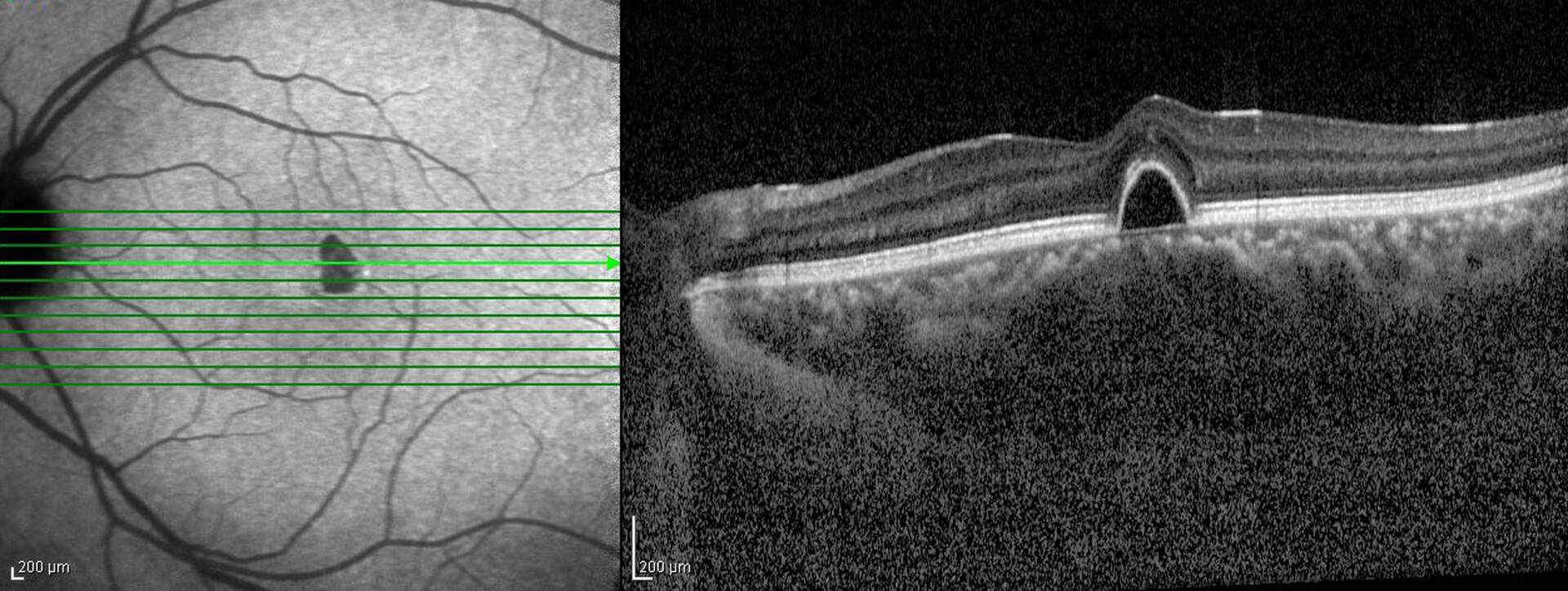
Optical coherence tomography works by using interferometry, enabling eye care professionals to image tissue with light instead of X-rays, sound or radio frequency. It works by shining a beam of light into the eye. Different tissues reflect part of this light, and images are built based on this internal reflection differential. 


An OCT images to approximately 2-3 mm below the surface of the tissue. Images are obtained clearly through a transparent window, such as the cornea. The light that emits into the eye is safe. The output is near-infrared light so that no eye damage will occur. 
What Happens During an OCT Scan?

An OCT may very well be the easiest medical imaging test you will ever take. The test only takes about 5 minutes to perform. With most OCT machines, you simply place your head in a headrest. Your optometrist will calibrate the instrument and instruct you to look at a light target inside the machine. The optometrist will then receive the image.


If your pupils are too small or the doctor wants to image a specific area, your doctor may use medicated eye drops to dilate your pupils. Most people do not require dilation.

Clinical Applications of Optical Coherence Tomography — Woy Woy, NSW — Peninsula Eyecare Izzards Optometrists

What Are OCT Scans Used For?

An OCT can be very helpful in the evaluation of many eye conditions, including:

Central serous retinopathy

Diabetic retinopathy

Glaucoma

Macular degeneration

Macular holes

Pre-retinal macular fibrosis (cellophane maculopathy)

An OCT represents some of the latest technological advances in eye care that are available at a minimal cost to the patient. 

Book an OCT Scan today at Woy Woy - 02 4341 2083 or Umina - 02 4342 1088.

Book an OCT Scan today at Woy Woy - 02 4341 2083 or Umina - 02 4342 1088.

Share by: